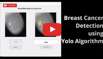
Breast Cancer Detection using Yolo Algorithm
Explore breast cancer detection using the YOLO algorithm for fast and accurate tumor identification. Learn how deep learning enhances medical image analysis for early diagnosis.
Project Methodology & Algorithm Details
Abstract
Introduction
Breast cancer is the most common worldwide tumor in the female population. Previous randomized trials and incidence-based mortality studies have demonstrated a sig-nificant reduction in breast cancer mortality associated with participation in breast screening programs. However, the problem of false positives and false negatives persists as a concern. Most of these errors can be attributed to dense breasts (masking effect), as well as human factors such as radiologist perception and erroneous decision-making behaviors. Additionally, the inherent imaging characteristics of tumors contribute to the issue, with benign masses often resembling malignant ones and malignant masses sometimes mimicking benign ones. During the breast cancer diag-nosis process, the physician aims to detect all the regions of interest (ROIs) in the whole mammogram: masses, calcifi-cations, distortions, etc. Detection in the early stage of the disease is critical for planning new examinations, therapies, or lines of intervention. A missed detection, on the other hand, may result in irreversible injury to the patient. For this reason, breast cancer detection is the most complicated but also the most important task. Unfortunately, several pro-posed solutions in the literature do not aim to analyze the entire image, but rather limit detection to patch classifica-tion: the ROIs are first manually selected and cropped, and then the classifiers are trained to distinguish the crops. How-ever, to support and imitate the physician’s diagnostic pro-cess, an architecture capable of detecting all ROIs within the whole mammogram is required. Faster R-CNN, RetinaNet, and Yolo have encouraged the development of systems for breast cancer detection. These frameworks certainly introduce two main difficulties:
(1) the models have to learn the features of the whole mammogram, and the image resiz-ing required for training may result in the loss of critical details;
(2) since the model has to detect all ROIs among all patches of healthy tissue (i.e., non-ROIs), an unavoidable increase in the error rate must be faced. However, Yolo has proven to be an excellent tool in numerous scenarios, achiev-ing higher accuracy and inference speed rates than its object detector competitors.
Need the Complete Source Code & Report for This Project?
Get fully documented code, dataset, and step-by-step guidance before your academic deadline.
Customize This Project
Direct review by PhD Lead Engineer
Related Project Demos


Need Custom Engineering Code?
Our PhD engineers provide complete MATLAB/Simulink projects with verified test cases and IEEE reports.
Order Project Solution →